Subjective:
The patient is a 22 y.o. male who presented to our office with a chief complaint of odontalgia in the left mandibular molar region.
Objective:
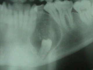
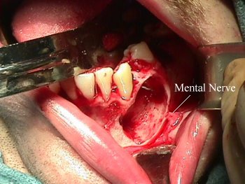
The patient has no allergies. There are no serious illnesses. Incidental panoramic radiographic findings revealed a left mandibular radiolucent lesion with associated impacted tooth # 21 and an over retained left mandibular first primary molar. The radiolucent lesion was well-delineated with calcific flecks. Roots of teeth #'s 20 and 22 were divergent. Tooth # 21 was at the inferior border of the mandible. Teeth #'s 1, 16, 17, and 32 were noted to be impacted. Carious lesions were noted in teeth #'s 2, 14, 15, 19, and 31. Tooth # 30 was missing and was extracted by his general dentist in 1997. Oral examination revealed a Class II malocclusion with anterior-posterior incisor discrepancy and anterior open-bite. Gingival erythema was diffuse. Expansion of the buccal cortical plate was noted, however no bruit or thrill was present. There was no paresthesia of the mental nerve.
Assessment:
- Dental Caries
- Class II Malocclusion with Apertognathia
- Impacted Teeth #'s 1, 16, 17, 21, and 32
- Over-retained Tooth # L
- Gingivitis
- Mandibular Odontogenic Cyst or Tumor
Plan:
- Out-patient General Anesthesia
- Aspiration of Lesion
- Extraction of Teeth #'s 1, 16, 17, 19, 21, 32, and L.
- Enucleation of Mandibular Lesion.
Results:
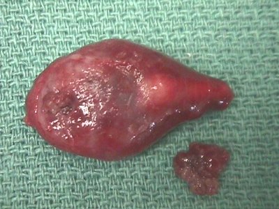
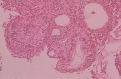
- Odontogenic Adenomatoid Tumor
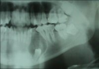
Radiolucent defect. | 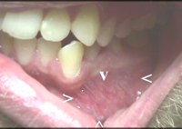
Expansion of buccal cortical plate. |
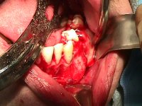
Exposed cortical plate expansion. | 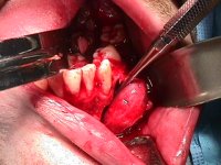
Enucleating lesion. |
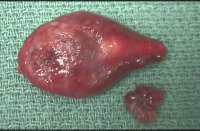
Tumor with material from lumen. | 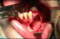
Osseous defect with intact mental nerve. |
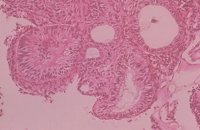
Odontogenic Adenomatoid Tumor |
Patient treated by:
- Dr. Steven R. Tucker Oral and Maxillofacial Surgery
 Steven R. Tucker DMD, PSC
 Oral and Maxillofacial Surgery
 909 Scherm Road
 Owensboro, KY 42301
 phone: 1-270-926-4107
 fax: 1-270-926-4166
 url: http://www.srt-psc.com

Last Modified: September 1, 1999