Subjective:
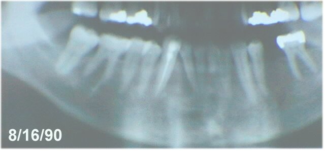
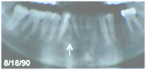
Patient was referred by her family dentist for evaluation of a radiolucent interradicular lesion in the mandible between teeth #'s 26 and 27. Tooth number 27 had recently had a root canal. Patient was asymptomatic.
Objective:
The patient is a healthy 68 y.o. white female. Oral examination revealed a Class I malocclusion, no mobility of teeth #'s 26 and 27. Tooth number 26 is vital. There was no expansion of the buccal or lingual cortical plate of the mandible. No bruit could be heard on auscultation of the mandible. No thrill was palpable. Preoperative aspiration of lesion was negative for a vascular lesion. Radiographic examination revealed an interradicular radiolucent defect between teeth #'s 26 and 27. The roots of teeth #'s 26 and 27 were noted to be divergent apically. Tooth number 27 had had a root canal.
 click on image
Assessment: (Preoperative Differential Diagnosis)
- Lateral Periodontal Cyst
- Odontogenic Keratocyst
- Giant Cell Granuloma
- Brown Tumor of Hyperparathyroidism
- Vascular Lesion (ruled out by aspiration)
- Ameloblastoma
- Benign Tumor of Periodontal Ligament Origin
Plan:
- IV sedation / local anesthesia
- Exploration / Enucleation of mandibular radiolucent lesion
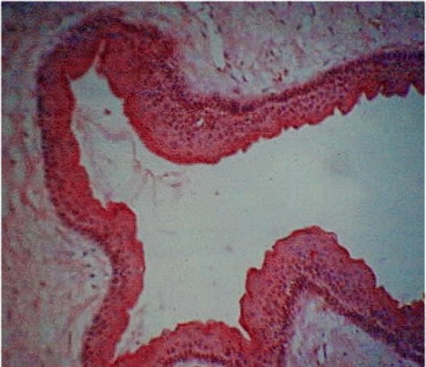
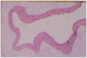
- Histopathologic examination of lesion by the University of Kentucky Oral Pathology Laboratory
 click on image
Diagnosis:
-
Odontogenic Keratocyst
Post Operative Follow-up
 click on image
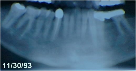
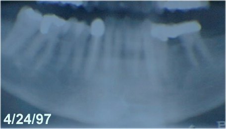
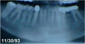
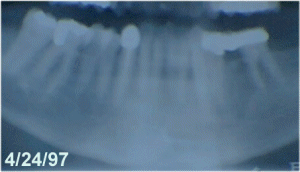
Note ossification of cystic defect and movement of roots back to original position.
- 09/06/90
- 09/26/91
- 10/15/92
- 11/30/93
- 01/03/95
- 04/04/96
- 04/24/97
 click on image
Patient treated by Dr. Steven R. Tucker
 Steven R. Tucker DMD, PSC
 Oral and Maxillofacial Surgery
 909 Scherm Road
 Owensboro, KY 42301
 1-502-926-4107
 fax: 1-502-926-4166

Last Modified: April 30, 1997