Subjective:
The patient is a 43 y.o.male injured by falling rocks in an underground coal mining accident. The patient was transferred, via ambulance, to Owensboro Mercy Health System from an adjacent county hospital. The patient was laying face down with a nasopharyngeal airway in place. The patient had no loss of consciousness, but had difficulty speaking and breathing when in a suspine position, secondary to having a flail maxilla and mandible.Objective:
The patient has no allergies. There are no serious illnesses and the patient takes no medications. The patient had been previously hospitalized for three separate mining accidents, resulting in blindness in his right eye, lumbar vertebrae fractures, and fracture of his left leg.The head was normocephalic. A scalp abrasion was present at the vertex. The patient had no vision in O.D., the pupil was irregular with a diameter of 7 mm. Bilateral periorbital edema and ecchymosis was noted. Epistaxis was present. The nasal complex and nasofrontal junction were intact. Teeth #s 24 and 25 were avulsed. Class III dental fractures of teeth #s 7, 26, and 31 were present. Partial avulsion of teeth #s 2, 15, and 16 was associated with dentoalveolar fractures of the maxilla, splitting the palate and creating right and left, anterior and posterior dentoalveolar segments. The palatal lacerations communicated with the maxillary vestibular lacerations and comminuted Le Fort I fracture creating traumatic oral antral openings. The split palate ran anteriorly between teeth #s 8 and 9, resulting in an oral nasal opening. Compound, comminuted right and left mandibular body fractures had associated through and through facial lacerations. A triangular segment of the left mandibular body was avulsed. Mental nerve anesthesia was present. Laceration of the lateral border of the tongue and multiple intraoral vestibular lacerations were noted.
Assessment:
- Comminuted Le Fort I Fracture with Four Dentoalveolar Segments
- Palatal Laceration
- Comminuted Palatal Fracture
- Compound, Comminuted Right Mandibular Body Fracture with Through and Through Facial Laceration
- Compound, Comminuted Left Mandibular Body Fracture with Through and Through Facial Laceration
- Right Mandibular Condyle Fracture
- Left Mandibular Condyle Fracture
- Right Mandibular Coronoid Process Fracture
- Right and Left Oral Antral Communications with Maxillary Vestibular Lacerations
- Oral Nasal Communication
- Avulsed Teeth #s 24 and 25
- Class III Dental Fractures Teeth #s 7, 26, and 31
- Partial Avulsion Teeth #s 2, 15, and 16
- Tongue Laceration
- Maxillary and Mandibular Vestibular Lacerations
Plan / Treatment:
- General Surgery Consultation for Tracheostomy
- Surgical Extraction of Teeth #s 2, 15, 16, 26, and 31
- Application of Maxillary and Mandibular Arch Bars
- Open Reduction Palatal Fractures
- Closure of Palatal Lacerations
- Open Reduction Le Fort I Fracture
- Closure of Tongue Laceration
- Closure of Maxillary and Mandibular Vestibular Lacerations
- Application of Intermaxillary Fixation
- Open Reduction Rigid Internal Fixation Left Mandibular Body Fracture
- Open Reduction Rigid Internal Fixation Right Mandibular Body Fracture
- Debridement and Closure of Facial Lacerations
- Closed Reduction Left Mandibular Condyle
- Open Reduction Rigid Internal Fixation Right Mandibular Condyle Fracture via Risdon Approach
- Removal of Fractured Coronoid Process
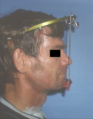
- Application of Joe Hall Morris Headframe
- Application of Joe Hall Morris Biphase to Mandibular Symphysis
- External Craniomandibular Fixation
- Future Root Canal Therapy Tooth # 7
- Future Maxillary and Mandibular Removal Partial Dentures
Results:
J. H. Morris headframe used in conjunction with Morris biphasic
external pin system.
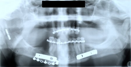
Panoramic radiograph after removal of headframe, biphase, and intermaxillary fixation.
Patient treated by:
- Dr. Steven R. Tucker Oral and Maxillofacial Surgery
 Steven R. Tucker DMD, PSC
 Oral and Maxillofacial Surgery
 909 Scherm Road
 Owensboro, KY 42301
 phone: 1-270-926-4107
 fax: 1-270-926-4166
 url: www.srt-psc.com
srt@srt-psc.com
Last Modified: October 31, 1999